
The problem
In day-to-day operations, scanning defects can delay specialist review. When the slide image, scanner metadata, barcode and quality protocol are scattered across different screens, documents or team records, problems do not become visible in time.
Fragmented data delays preparation of the slide quality review queue and forces staff to search manually for supporting evidence again. Before making a decision, the pathology technician and pathologist have to complete missing fields and verify inconsistencies one by one.
A common exception is that a valid specimen with limited material may fall below standard coverage. Incomplete or incorrect automation that overlooks this context could lead to a valid specimen being rescanned or an inadequate scan being accepted, so the output cannot be applied directly.
What we set out to improve
- Rate of confirmed rescan candidates
- Rate of outputs corrected or rejected by the pathology technician and pathologist
- Time between the source record and the review output
How the system works
- 01Define the scope of the review and the authorised pathology technician and pathologist roles
- 02Collect the inputs: slide image, scanner metadata, barcode and quality protocol
- 03Check source identity, date, version and data freshness
- 04Check barcode and file integrity
- 05Classify focus and coverage signals
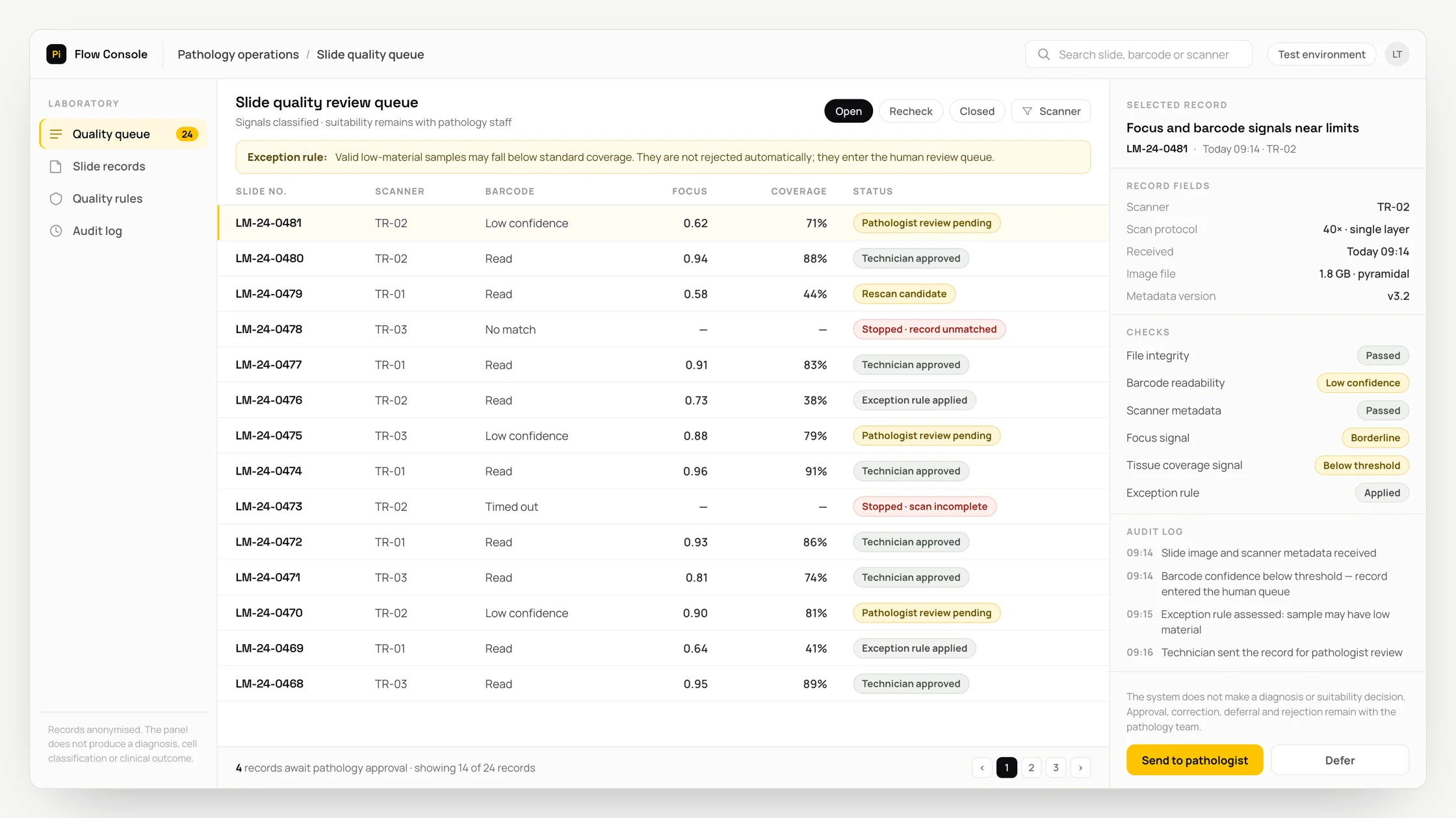
- 06Check the exception: a valid specimen with limited material may fall below standard coverage
- 07Place low-confidence or inconsistent records in a separate human review queue
- 08Write the source identity, rule, output and time to the audit trail
- 09Have the pathology technician and pathologist approve, correct, defer or reject the output
Methods we used
- A technical image quality model that does not classify pathological findings
- Source, format, version and required-field checks for the slide image, scanner metadata, barcode and quality protocol
- A missing-data warning instead of an estimate when inputs are missing or inconsistent
- Checking exceptions separately from the main rule
- Storing the source identity, rule result, slide quality review queue and human decision together
Where people stay involved
The system stops after preparing the slide quality review queue. The pathology technician and pathologist review the sources and the exception, then approve, correct or reject the output. Direct application is disabled because it could lead to a valid specimen being rescanned or an inadequate scan being accepted; the system does not replace specialist judgement.
Data and security
Access to the slide image, scanner metadata, barcode and quality protocol is limited to the minimum permissions required for each role. Identifiers and sensitive fields are masked where possible, and only the necessary section is sent to the model rather than the entire raw file. Access, outputs and decisions by the pathology technician and pathologist are recorded; the system does not enter production until the organisation has approved the retention period and data location.
Who this suits
A good fit
- Digital pathology laboratories
Not a good fit
- Processes seeking clinical interpretations
FAQ
Frequently asked questions
Which inputs are used?
The system uses the slide image, scanner metadata, barcode and quality protocol. If a required field is unavailable, it does not produce a definitive result and presents the missing source for review by the pathology technician and pathologist.
At what point does a person make the decision?
The workflow stops when the slide quality review queue is ready and waits for approval from the pathology technician and pathologist. The specialists may correct, defer or reject the output and record their reasons.
Can the output be audited?
The system is designed to store the source identity, applied rule, exception, output and human decision together. This makes it possible to review later how the source data led to a particular recommendation.
Project led by: DijitalPi
✦ PI ASSISTANT · ARTIFICIAL INTELLIGENCE
onlineAsk the DijitalPi AI Assistant your question
No need to fill in a form and wait for a reply — write your question and let Pi, trained on DijitalPi's 20 years of know-how, answer within seconds.
- Instant answers, 24/7
- Service-specific context — no generic replies
- Can connect you to a free consultation if you wish
Pi Assistant answers are for information purposes; for a proposal get in touch.
ASK AI ABOUT DIJITALPI
Let an AI explain what DijitalPi does.
Opens your chosen assistant with a ready research prompt. It reads the site live and answers.
 Cookie preferences
We use cookies to improve your experience and to analyse site traffic anonymously.Details: Cookie Policy · Privacy